Workshop – Technology Supporting Precision Medicine
- Posted by Stefano Pipi
- Categories Seminars
- Date 30 Novembre 2021

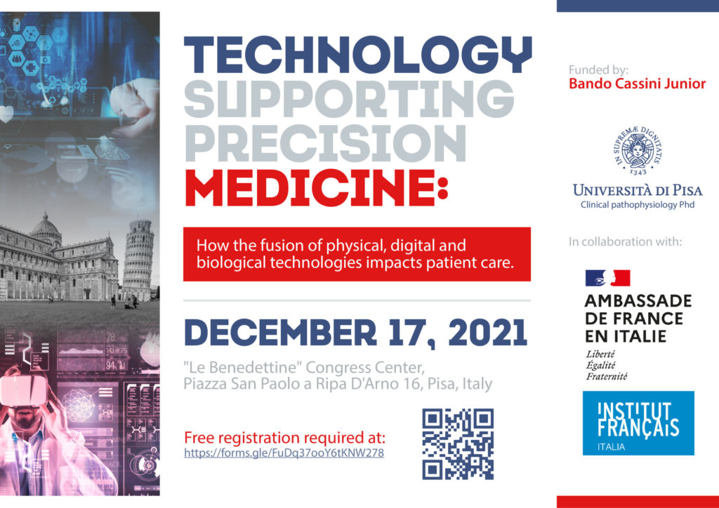
December 17th will be held the workshop “Technology supporting precision medicine: how the fusion of physical, digital and biological technologies impacts patient care.”
The workshop is organized by dr. Greta Barbieri in collaboration with the University of Pisa and Ambassade De France en Italie together with the Institut Français – Italia.
To partecipate in the event, you can register at the following link: https://forms.gle/FuDq37ooY6tKNW278
Cassini Program:
The initiative aims to allow PhD students and scholars from Italian Universities
to create or develop scientific relationships with French Universities and research centers. Cassini Junior (reserved for PhD students) and Cassini Senior (reserved for researchers) provide the allocation of founding for the organization of a scientific event in connection with the French and Italian cultural areas.
Scientific rationale:
Recent challenges to which the health system has been subjected, require reflection on the need to implement medicine traditional tools with modern methods that draw on multiple scientific fields. The goal of the physician today is no longer just the pathology, but the individual, with a view to personalized medicine. This cultural transition is not achievable except with integration of high-level technological tools in clinical practice.
Ultrasound (US) is a method well known for its feasibility and high precision for assessing dynamic and temporal changes. In fact, it can be used at bedside and provide fundamental information for diagnosis, monitoring and prognostic evaluation in cardiovascular diseases. In the context of multiparametric evaluations, US could allow a “personalization” of therapy in the context of cardiovascular pathologies in different hemodynamic settings.
Very recently, lung US has been shown to play a crucial role in COVID-19 pandemic, providing an easily available and quick to use tool in a context of logistical difficulty, usable in different settings for diagnosis and identification of patient’s suitable path.
While research in the ‘-omic’ sciences has resulted in improved understanding of the relationships between genes, proteins and disease, recently, the approach of precision medicine has been extended to medical imaging. The so-called “radiomics” allows to obtain quantitative parameters from spatial data sets, to create large data sets with the aim of quantifying morphological characteristics, impossible to understand by visual evaluation. This has proven to be a valuable tool in oncology and is also increasingly used in cardiovascular medicine.
Precision medicine needs of accurate diagnostic tools. Applications of 7T MR imaging in patients with neurological disorders having not conclusive conventional imaging could open new perspective in the era of precision medicine.
Dual-energy CT (DECT) may allow for better tissue characterization and therefore enhanced visualization of heart and vessels. Given the unique feature of allowing the differentiation of iodine attenuation characteristics when exposed to different photon energy levels, DECT allows mapping of iodine distribution.
Ultra-high magnetic field MR systems (7T) are research instruments developed to deeply explore pathologic processes in individual patients.
The multidistrict imaging assessment provide large sets of measurements. Therefore, novel big data analytic approaches may be well-suited for data mining and for pattern recognition
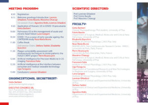
Event brochure – Technology supporting medicine


